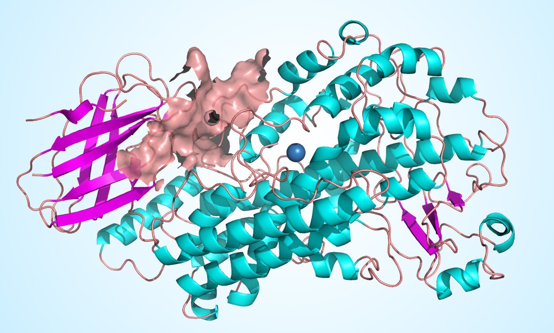
At the University of Innsbruck, a pharmacy team has been inquiring about normal items for their anti-inflammatory impacts for quite a while. The investigations will be done together with a worldwide consortium with the cooperation of scientists from Germany, France, and Italy. In these investigations, information rose that indicated vitamin E and related structures as an on-screen character in the inflammatory procedure.
Blog Archives
Induction of peroxisomal changes in oligodendrocytes treated with 7-ketocholesterol: Attenuation by α-tocopherol
Nury T, Sghaier R, Zarrouk A, Ménétrier F, Uzun T, Leoni V, Caccia C, Meddeb W, Namsi A, Sassi K, Mihoubi W, Riedinger JM, Cherkaoui-Malki M, Moreau T, Vejux A, Lizard G
Biochimie. 2018 Oct;153:181-202. doi: 10.1016/j.biochi.2018.07.009. Epub 2018 Jul 19.
Abstract
The involvement of organelles in cell death is well established especially for endoplasmic reticulum, lysosomes and mitochondria. However, the role of the peroxisome is not well known, though peroxisomal dysfunction favors a rupture of redox equilibrium. To study the role of peroxisomes in cell death, 158 N murine oligodendrocytes were treated with 7-ketocholesterol (7 KC: 25-50 μM, 24 h). The highest concentration is known to induce oxiapoptophagy (OXIdative stress + APOPTOsis + autoPHAGY), whereas the lowest concentration does not induce cell death. In those conditions (with 7 KC: 50 μM) morphological, topographical and functional peroxisome alterations associated with modifications of the cytoplasmic distribution of mitochondria, with mitochondrial dysfunction (loss of transmembrane mitochondrial potential, decreased level of cardiolipins) and oxidative stress were observed: presence of peroxisomes with abnormal sizes and shapes similar to those observed in Zellweger fibroblasts, lower cellular level of ABCD3, used as a marker of peroxisomal mass, measured by flow cytometry, lower mRNA and protein levels (measured by RT-qPCR and western blotting) of ABCD1 and ABCD3 (two ATP-dependent peroxisomal transporters), and of ACOX1 and MFP2 enzymes, and lower mRNA level of DHAPAT, involved in peroxisomal β-oxidation and plasmalogen synthesis, respectively, and increased levels of very long chain fatty acids (VLCFA: C24:0, C24:1, C26:0 and C26:1, quantified by gas chromatography coupled with mass spectrometry) metabolized by peroxisomal β-oxidation. In the presence of 7 KC (25 μM), slight mitochondrial dysfunction and oxidative stress were found, and no induction of apoptosis was detected; however, modifications of the cytoplasmic distribution of mitochondria and clusters of mitochondria were detected. The peroxisomal alterations observed with 7 KC (25 μM) were similar to those with 7 KC (50 μM). In addition, data obtained by transmission electron microcopy and immunofluorescence microscopy by dual staining with antibodies raised against p62, involved in autophagy, and ABCD3, support that 7 KC (25-50 μM) induces pexophagy. 7 KC (25-50 μM)-induced side effects were attenuated by α-tocopherol but not by α-tocotrienol, whereas the anti-oxidant properties of these molecules determined with the FRAP assay were in the same range. These data provide evidences that 7 KC, at concentrations inducing or not cell death, triggers morphological, topographical and functional peroxisomal alterations associated with minor or major mitochondrial changes.
δ-Tocotrienol, Isolated from Rice Bran, Exerts an Anti-Inflammatory Effect via MAPKs and PPARs Signaling Pathways in Lipopolysaccharide-Stimulated Macrophages
Shen J, Yang T, Xu Y, Luo Y, Zhong X, Shi L, Hu T, Guo T, Nie Y, Luo F, Lin Q
Int J Mol Sci. 2018 Oct 4;19(10). pii: E3022. doi: 10.3390/ijms19103022.
Abstract
δ-Tocotrienol, an important component of vitamin E, has been reported to possess some physiological functions, such as anticancer and anti-inflammation, however their molecular mechanisms are not clear. In this study, δ-tocotrienol was isolated and purified from rice bran. The anti-inflammatory effect and mechanism of δ-tocotrienol against lipopolysaccharides (LPS) activated pro-inflammatory mediator expressions in RAW264.7 cells were investigated. Results showed that δ-tocotrienol significantly inhibited LPS-stimulated nitric oxide (NO) and proinflammatory cytokine (TNF-α, IFN-γ, IL-1β and IL-6) production and blocked the phosphorylation of c-Jun N-terminal kinase (JNK) and extracellular regulated protein kinases 1/2 (ERK1/2). δ-Tocotrienol repressed the transcriptional activations and translocations of nuclear factor-kappa B (NF-κB) and activator protein-1 (AP-1), which were closely related with downregulated cytokine expressions. Meanwhile, δ-tocotrienol also affected the PPAR signal pathway and exerted an anti-inflammatory effect. Taken together, our data showed that δ-tocotrienolinhibited inflammation via mitogen-activated protein kinase (MAPK) and peroxisome proliferator-activated receptor (PPAR) signalings in LPS-stimulated macrophages.
The Effect of Magnesium and Vitamin E Co-Supplementation on Glycemic Control and Markers of Cardio-Metabolic Risk in Women with Polycystic Ovary Syndrome: A Randomized, Double-Blind, Placebo-Controlled Trial
Jamilian M, Sabzevar NK, Asemi Z
Horm Metab Res. 2018 Oct 4. doi: 10.1055/a-0749-6431. [Epub ahead of print]
Abstract
Data on the effects of magnesium and vitamin E co-supplementation on glycemic control and markers of cardio-metabolic risk of patients with polycystic ovary syndrome (PCOS) were collected. This investigation was conducted to evaluate the effects of magnesium and vitamin E co-supplementation on glycemic control and markers of cardio-metabolic risk in women with PCOS. This randomized, double-blind, placebo-controlled trial was carried out on 60 women with PCOS, aged 18-40 years old. Participants were randomly divided into two groups to receive 250 mg/day magnesium plus 400 mg/day vitamin E supplements or placebo (n=30 each group) for 12 weeks. Fasting blood samples were taken at baseline and after the 12-week intervention to quantify related variables. After the 12-week intervention, compared with the placebo, magnesium and vitamin E co-supplementation led to a significant reduction in serum insulin levels (-1.1±3.0 vs. +1.6±3.7 μIU/ml, p=0.003) and homeostatic model of assessment for insulin resistance (-0.2±0.7 vs. +0.4±0.9, p=0.002), and a significant increase in the quantitative insulin sensitivity check index (+0.01±0.01 vs. -0.009±0.02, p=0.003). Furthermore, magnesium plus vitamin E supplementation significantly decreased serum triglycerides (-15.0±24.4 vs. +6.7±22.2 mg/dl, p=0.001) and VLDL-cholesterol concentrations (-3.0±4.9 vs. +0.6±2.4 mg/dl, P=0.01) compared with the placebo. A trend toward a greater decrease in total cholesterol levels was observed in magnesium plus vitamin Egroup compared to placebo group (-7.0±32.6 vs. +8.1±26.6 mg/dl, p=0.05). In conclusion, magnesium and vitamin E co-supplementation for 12 weeks to PCOS women had beneficial effects on parameters of insulin metabolism and few markers of cardio-metabolic risk.
Why vitamin E effect is often a matter of luck until now
Vitamin E’s positive effects often fail to manifest themselves as strongly as expected, but sometimes administering vitamin E actually has detrimental effects. An international team has now found a possible cause for this. It has shown that the effect of vitamin E, which is taken as a tablet or capsule, is not based on the vitamin itself, but rather on the effect of a metabolite. This socalled alpha-carboxychromanol has e. g. a promising anti-inflammatory effect.
Nonalcoholic steatohepatitis, obesity, and cardiac dysfunction
Mathews SE, Kumar RB, Shukla AP
Curr Opin Endocrinol Diabetes Obes. 2018 Oct;25(5):315-320. doi: 10.1097/MED.0000000000000432.
Abstract
PURPOSE OF REVIEW:
Obesity and nonalcoholic steatohepatitis (NASH) are epidemiologically and pathophysiologically linked disorders. Here, we summarize the effect of obesity on NASH and how it has a cascading effect on cardiovascular dysfunction. We also review the current and emerging treatment options for NASH.
RECENT FINDINGS:
The link between NASH and cardiac dysfunction has been further delineated in recent studies demonstrating endothelial dysfunction, diastolic dysfunction, and increased coronary artery calcification in patients with known NASH. Standard treatment of obesity with lifestyle interventions including diet, exercise, and behavioral modification has been shown to improve NASH as well as reduce cardiovascular dysfunction. In addition to FDA-approved drugs like vitamin E and pioglitazone, several agents including NGM282, obeticholic acid, elafibranor, and liraglutide are currently being investigated for their therapeutic potential in NASH. Recent studies show that bariatric surgery results in significant improvement or resolution of NASH.
SUMMARY:
Obesity is a major factor in the development of nonalcoholic fatty liver disease (NAFLD) and its progression to steatohepatitis. Patients with NAFLD have a significant increase in cardiovascular disease risk. For biopsy-proven NASH, vitamin E and pioglitazone are the recommended medical treatments in addition to lifestyle modification.
Vitamin E: Sources, Benefits & Risks
Vitamin E is a vital nutrient for good health, and it’s found in a wide variety of foods and supplements. The best way to consume this vitamin is through a healthy diet. Deficiency is rare, and overdosing by using supplements is a concern. Those who have certain health conditions or take certain medicines should be cautious with supplements.
Safety Assessment of Tocopherols and Tocotrienols as Used in Cosmetics
Fiume MM, Bergfeld WF, Belsito DV, Hill RA, Klaassen CD, Liebler DC, Marks JG Jr, Shank RC, Slaga TJ, Snyder PW, Andersen FA, Heldreth B
Send to Int J Toxicol. 2018 Sep/Oct;37(2_suppl):61S-94S. doi: 10.1177/1091581818794455.
Abstract
The Cosmetic Ingredient Review (CIR) Expert Panel (Panel) assessed the safety of 14 tocopherols and tocotrienols and concluded these ingredients are safe as used in cosmetics. The tocopherols are reported to function in cosmetics as antioxidants or skin-conditioning agents; in contrast, tocotrienols are not reported to function as an antioxidants in cosmetics but as a light stabilizer, oral care agent, or skin-conditioning agent. The Panel reviewed the new and existing animal and clinical data to determine the safety of these ingredients and found it appropriate to extrapolate the existing information to conclude on the safety of all the tocopherols and tocotrienols.
The Role of Vitamin E in the Treatment of NAFLD
Perumpail BJ, Li AA, John N, Sallam S, Shah ND, Kwong W, Cholankeril G, Kim D, Ahmed A
Diseases. 2018 Sep 24;6(4). pii: E86. doi: 10.3390/diseases6040086
Abstract
There has been a growing interest in the role of vitamin E supplementation in the treatment and/or prevention of nonalcoholic fatty liver (NAFLD). We performed a systematic review of the medical literature from inception through 15 June 2018 by utilizing PubMed and searching for key terms such as NAFLD, vitamin E, alpha-tocopherol, and nonalcoholic steatohepatitis (NASH). Data from studies and medical literature focusing on the role of vitamin E therapy in patients with NAFLD and nonalcoholic steatohepatitis (NASH) were reviewed. Most studies assessing the impact of vitamin E in NAFLD were designed to evaluate patients with NASH with documented biochemical and histological abnormalities. These studies demonstrated improvement in biochemical profiles, with a decline in or normalization of liver enzymes. Furthermore, histological assessment showed favorable outcomes in lobular inflammation and hepatic steatosis following treatment with vitamin E. Current guidelines regarding the use of vitamin E in the setting of NAFLD recommend that vitamin E-based treatment be restricted to biopsy-proven nondiabetic patients with NASH only. However, some concerns have been raised regarding the use of vitamin E in patients with NASH due to its adverse effects profile and lack of significant improvement in hepatic fibrosis. In conclusion, the antioxidant, anti-inflammatory, and anti-apoptotic properties of vitamin E accompanied by ease-of-use and exceptional tolerability have made vitamin E a pragmatic therapeutic choice in non-diabetic patients with histologic evidence of NASH. Future clinical trials with study design to assess vitamin E in combination with other anti-fibrotic agents may yield an additive or synergistic therapeutic effect.
Antiglycation study of HMG-R inhibitors and tocotrienol against glycated BSA and LDL: A comparative study.
Nabi R, Alvi SS, Khan RH, Ahmad S, Ahmad S, Khan MS
Int J Biol Macromol. 2018 Sep;116:983-992. doi: 10.1016/j.ijbiomac.2018.05.115. Epub 2018 May 18.
Abstract
Non-enzymatic glycation mediated advanced glycation end products (AGEs) generation results in the pathogenesis of diabetic complications and atherosclerotic cardiovascular disease (ASCVD) which is greatly influenced by 3-hydroxy-3-methyl-glutaryl Co-A reductase (HMG-R) activity. HMG-R inhibitors, statins, are well known for reducing mortality and morbidity of ASCVD in patients with diabetes due to their pleiotropic effects independent of cholesterol lowering. Due to distinct chemical structures, various statins may play important role in the inhibition of AGEs mediated pathologies. Herein, we evaluated the anti-glycating potential of atorvastatin (AT), rosuvastatin (RT), pitavastatin (PT), fluvastatin (FT), simvastatin (ST) alone as well in combination with ezetimibe (EZ) and tocotrienol (TT) against d-ribose mediated BSA and LDL glycation by various physicochemical approaches. Our data suggested that AT, TT, RT, EZ, EZ-AT, and EZ-RT were able to substantially inhibit the AGEs formation via modulation of hyperchromicity, fluorogenic AGEs, % contribution of α-helix and β-sheets to protein secondary structure, amide-I band stretching, carbonyl and HMF content in Gly-BSA as well as Gly-LDL. On the basis of above findings, we concluded that HMG-R inhibitors and TT, alone or in combination with EZ, may be established as terrific therapeutic agents for the patients suffering from AGEs induced diabetic cum ASCVD complications.