Tocotrienol supplementation suppressed bone resorption and oxidative stress in postmenopausal osteopenic women: a 12-week randomized double-blinded placebo-controlled trial.

Shen CL, Yang S, Tomison MD, Romero AW, Felton CK, Mo H

Osteoporos Int. 2018 Jan 12. doi: 10.1007/s00198-017-4356-x. [Epub ahead of print]

Abstract

Tocotrienols have shown bone-protective effect in animals. This study showed that a 12-week tocotrienol supplementation decreased concentrations of bone resorption biomarker and bone remodeling regulators via suppressing oxidative stress in postmenopausal osteopenic women.

INTRODUCTION:

Tocotrienols (TT) have been shown to benefit bone health in ovariectomized animals, a model of postmenopausal women. The purpose of this study was to evaluate the effect of 12-week TT supplementation on bone markers (serum bone-specific alkaline phosphatase (BALP), urine N-terminal telopeptide (NTX), serum soluble receptor activator of nuclear factor-kappaB ligand (sRANKL), and serum osteoprotegerin (OPG)), urine calcium, and an oxidative stress biomarker (8-hydroxy-2′-deoxyguanosine (8-OHdG)) in postmenopausal women with osteopenia.

METHODS:

Eighty-nine postmenopausal osteopenic women (59.7 ± 6.8 year, BMI 28.7 ± 5.7 kg/m2) were randomly assigned to three groups: (1) placebo (430 mg olive oil/day), (2) low TT (430 mg TT/day, 70% purity), and (3) high TT (860 mg TT/day, 70% purity). TT, an extract from annatto seed with 70% purity, consisted of 90% delta-TT and 10% gamma-TT. Overnight fasting blood and urine samples were collected at baseline, 6, and 12 weeks for biomarker analyses. Eighty-seven subjects completed the 12-week study.

RESULTS:

Relative to the placebo group, there were marginal decreases in serum BALP level in the TT-supplemented groups over the 12-week study period. Significant decreases in urine NTX levels, serum sRANKL, sRANKL/OPG ratio, and urine 8-OHdG concentrations and a significant increase in BALP/NTX ratio due to TT supplementation were observed. TT supplementation did not affect serum OPG concentrations or urine calcium levels throughout the study period. There were no significant differences in NTX level, BALP/NTX ratio, sRANKL level, and sRANKL/OPG ratio between low TT and high TT groups.

CONCLUSIONS:

Twelve-week annatto-extracted TT supplementation decreased bone resorption and improved bone turnover rate via suppressing bone remodeling regulators in postmenopausal women with osteopenia. Such osteoprotective TT’s effects may be, in part, mediated by an inhibition of oxidative stress.

Read More

Vitamin E inhibits the UVAI induction of “light” and “dark” cyclobutane pyrimidine dimers, and oxidatively generated DNA damage, in keratinocytes

Delinasios GJ, Karbaschi M, Cooke MS, Young AR

Sci Rep. 2018 Jan 11;8(1):423. doi: 10.1038/s41598-017-18924-4.

Abstract

Solar ultraviolet radiation (UVR)-induced DNA damage has acute, and long-term adverse effects in the skin. This damage arises directly by absorption of UVR, and indirectly via photosensitization reactions. The aim of the present study was to assess the effects of vitamin E on UVAI-induced DNA damage in keratinocytes in vitro. Incubation with vitamin E before UVAI exposure decreased the formation of oxidized purines (with a decrease in intracellular oxidizing species), and cyclobutane pyrimidine dimers (CPD). A possible sunscreening effect was excluded when similar results were obtained following vitamin E addition after UVAI exposure. Our data showed that DNA damage by UVA-induced photosensitization reactions can be inhibited by the introduction of vitamin E either pre- or post-irradiation, for both oxidized purines and CPD (including so-called “dark” CPDs). These data validate the evidence that some CPD are induced by UVAI initially via photosensitization, and some via chemoexcitation, and support the evidence that vitamin E can intervene in this pathway to prevent CPD formation in keratinocytes. We propose the inclusion of similar agents into topical sunscreens and aftersun preparations which, for the latter in particular, represents a means to mitigate on-going DNA damage formation, even after sun exposure has ended.

Read More

Ischemia-Reperfusion Injury of Sciatic Nerve in Rats: Protective Role of Combination of Vitamin C with E and Tissue Plasminogen Activator

Apostolopoulou K, Konstantinou D, Alataki R, Papapostolou I, Zisimopoulos D, Kalaitzopoulou E, Bravou V, Lilis I, Angelatou F, Papadaki H, Georgiou CD, Chroni E

Neurochem Res. 2018 Jan 11. doi: 10.1007/s11064-017-2465-8. [Epub ahead of print]

Abstract

An ischemia/reperfusion injury of rat’s sciatic nerve was experimentally developed. In this model, we measured the in vivo production of superoxide radical, as a marker of oxidative stress and the occludin expression as an indicator of blood-nerve barrier function and we examined potential protective innervations against these abnormalities. Right sciatic nerves of the animals underwent 3 h of ischemia followed by 7 days of reperfusion and were divided into three groups: ischemic, pretreated with vitamin C in conjunction with vitamin E and treated with tissue plasminogen activator. Compared to measurements from left sciatic nerves used as sham, the ischemic group showed significantly increased superoxide radical and reduced expression of occludin in western blot and immunohistochemistry. No such differences were detected between sham and nerves in the vitamin or tissue plasminogen activator groups. It is suggested that the experimental ischemia/reperfusion model was suitable for studying the relationship between oxidative state and blood-nerve barrier. The reversion of abnormalities by the applied neuroprotective agents might prove to be a clinically important finding in view of the implication of vascular supply derangement in various neuropathies in humans.

Read More

The use of 99mTc-phytate for assessment the protective effect of vitamin E against hepatotoxicity induced by methotrexat in rat

Amirfakhrian H, Abedi SM, Sadeghi H, Azizi S, Hosseinimehr SJ

Nucl Med Rev Cent East Eur. 2018 Jan 10. doi: 10.5603/NMR.a2018.0006. [Epub ahead of print]

Abstract

In this study, we investigated the protective effect of vitamin E against methotrexate (MTX)-induced hepatotoxicity by quantitative liver 99mTc-phytate uptake and liver imaging and to compare its effect with histopathology in rat. Rats were divided into five groups as control, solvent, Vit E (100 mg/kg), MTX (20 mg/kg), Vit E + MTX and. Vit E was intraperitoneally administrated for 17 days before MTX injection and continued for 4 days. 99mTc-phytate was injected through the tail of rats after the drug administration. The percentage of the injected dose per gram of liver and spleen tissues (%ID/g) was calculated. Liver imaging was obtained with gamma camera. In other experiment, liver of treated rats were assessed for histopathology. 99mTc-phytate uptake per gram tissue of the livers as %ID/g in control, solvent, MTX, Vit E, Vit E + MTX and MTX groups were 8.99%  1.37, 8.53%  2.91, 8.65%  3.84, 3.22%  1.09 and 8.38%  2.68. Vit E administration with MTX resulted in a significant increasing in the level of %ID/g. Vit E treatment improved the shape of live in planner image. Histophatological examinations showed a protective effect of Vit E against MTX-induced hepatoxicity in rats. The results showed that Vit E significantly attenuates the MTX-induced hepatotoxicity in rats, and 99mTc-phytate uptake in liver as well as liver image to be acceptable techniques for assessment of liver and spleen damages and/or their tissues protective effects in animal model.

Read More

Health Benefits of Vitamin E

Vitamin E, a fat-soluble antioxidant, can only be obtained as a food supplement, but has widely-known health benefits for the skin, heart and brain. Deficiency of vitamin E is rarely naturally-occurring, but when it does appear, it is typically caused by fat malabsorption disorders or genetic abnormalities. Vitamin E is well-known in the cosmetic world for its skin benefits, but also protects against toxins that can deteriorate the eyes and brain.

Read More

Vitamin transporters in mice brain with aging

Marcos P, González-Fuentes J, Castro-Vázquez L, Lozano MV, Santander-Ortega MJ, Rodríguez-Robledo V, Villaseca-González N, Arroyo-Jiménez MM

J Anat. 2018 Jan 8. doi: 10.1111/joa.12769. [Epub ahead of print]

Abstract

Its high metabolic rate and high polyunsaturated fatty acid content make the brain very sensitive to oxidative damage. In the brain, neuronal metabolism occurs at a very high rate and generates considerable amounts of reactive oxygen species and free radicals, which accumulate inside neurons, leading to altered cellular homeostasis and integrity and eventually irreversible damage and cell death. A misbalance in redox metabolism and the subsequent neurodegeneration increase throughout the course of normal aging, leading to several age-related changes in learning and memory as well as motor functions. The neuroprotective function of antioxidants is crucial to maintain good brain homeostasis and adequate neuronal functions. Vitamins E and C are two important antioxidants that are taken up by brain cells via the specific carriers αTTP and SVCT2, respectively. The aim of this study was to use immunohistochemistry to determine the distribution pattern of these vitamin transporters in the brain in a mouse model that shows fewer signs of brain aging and a higher resistance to oxidative damage. Both carriers were distributed widely throughout the entire brain in a pattern that remained similar in 4-, 12-, 18- and 24-month-old mice. In general, αTTP and SVCT2 were located in the same regions, but they seemed to have complementary distribution patterns. Double-labeled cell bodies were detected only in the inferior colliculus, entorhinal cortex, dorsal subiculum, and several cortical areas. In addition, the presence of αTTP and SVCT2 in neurons was analyzed using double immunohistochemistry for NeuN and the results showed that αTTP but not SVCT2 was present in Bergmann’s glia. The presence of these transporters in brain regions implicated in learning, memory and motor control provides an anatomical basis that may explain the higher resistance of this animal model to brain oxidative stress, which is associated with better motor performance and learning abilities in old age.

Read More

Annatto: Delivering Tocotrienols from Amazonia

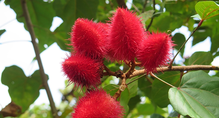
Today, annatto is known to be one of the superior sources of tocotrienols, whose researched health benefits mirror some of those passed down from ancient traditions. Unique among the plant kingdom, annatto produces only tocotrienols, whereas all other known sources of this vitamin E nutrient, such as palm and rice, deliver mixtures of tocopherols and tocotrienols, typically containing anywhere from 25-50% alpha-tocopherol. This is one ancient secret steeped into an Amazonian past.

Read More

Her son’s eczema inspired her to create an anti-inflammatory moisturising cream

Welcoming your first child is a time of great joy, but also a time of great anxiety. Worries about whether you’re feeding him right, carrying her correctly, bathing him properly and interpreting her heart-rending cries accurately, are common emotional companions for first-time parents. So, it’s hard enough coping with a healthy baby, but what if he or she also has a health problem?

Universiti Putra Malaysia (UPM) Department of Bioprocess Technology professor Dr Lai Oi Ming had her first, and only, child in 2013. But her joy in welcoming her firstborn was marred by his unexpected skin condition.

Read More

Vitamin E (α‑tocopherol) ameliorates aristolochic acid‑induced renal tubular epithelial cell death by attenuating oxidative stress and caspase‑3 activation.

Wu TK, Pan YR, Wang HF, Wei CW, Yu YL

Mol Med Rep. 2018 Jan;17(1):31-36. doi: 10.3892/mmr.2017.7921. Epub 2017 Oct 27.

Abstract

Aristolochic acid (AA) is a component identified in traditional Chinese remedies for the treatment of arthritic pain, coughs and gastrointestinal symptoms. However, previous studies have indicated that AA can induce oxidative stress in renal cells leading to nephropathy. α‑tocopherol exists in numerous types of food, such as nuts, and belongs to the vitamin E isoform family. It possesses antioxidant activities and has been used previously for clinical applications. Therefore, the aim of the present study was to determine whether α‑tocopherol could reduce AA‑induced oxidative stress and renal cell cytotoxicity, determined by cell survival rate, reactive oxygen species detection and apoptotic features. The results indicated that AA markedly induced H2O2 levels and caspase‑3 activity in renal tubular epithelial cells. Notably, the presence of α‑tocopherol inhibited AA‑induced H2O2 and caspase‑3 activity. The present study demonstrated that antioxidant mechanisms of α‑tocopherol may be involved in the increased survival rates from AA‑induced cell injury.

Read More

Vitamin E can improve behavioral tests impairment, cell loss, and dendrite changes in rats’ medial prefrontal cortex induced by acceptable daily dose of aspartame.

Rafati A, Noorafshan A, Jahangir M, Hosseini L, Karbalay-Doust S

Acta Histochem. 2018 Jan;120(1):46-55. doi: 10.1016/j.acthis.2017.11.004. Epub 2017 Nov 21.

Abstract

Aspartame is an artificial sweetener used in about 6000 sugar-free products. Aspartame consumption could be associated with various neurological disorders. This study aimed to evaluate the effect of aspartame onmedial Prefrontal Cortex (mPFC) as well as neuroprotective effects of vitamin E. The rats were divided into seven groups, including distilled water, corn oil, vitamin E (100mg/kg/day), and low (acceptable daily dose) and high doses of aspartame (40 and 200mg/kg/day) respectively, with or without vitamin E consumption, for 8 weeks. Behavioral tests were recorded and the brain was prepared for stereological assessments. Novel objects test and eight-arm radial maze showed impairmentoflong- and short-termmemoriesin aspartame groups. Besides, mPFC volume, infralimbic volume, neurons number, glial cells number, dendrites length per neuron,and number of spines per dendrite length were decreased by 7-61% in the rats treated with aspartame. However, neurons’ number, glial cells number, and rats’ performance in eight-arm radial mazes were improved by concomitant consumption of vitamin E and aspartame. Yet, the mPFC volume and infralimbic cortex were protected only in the rats receiving the low dose of aspartame+vitamin E. On the other hand, dendrites length, spines number,and novel object recognition were not protected by treatment with vitamin E+aspartame. The acceptable daily dose or higher doses of aspartame could induce memory impairments and cortical cells loss in mPFC. However, vitamin E could ameliorate some of these changes.

Read More